Orbital Tumors - Hemangiopericytoma
Description of Orbital Tumors - Hemangiopericytoma
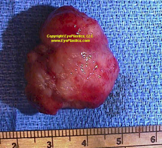
- Contractile cells that wrap around the endothelial cells of blood vessels are known as pericytes. Orbital hemangiopericytoma is a rare, solid, slow-growing tumour that arises from the proliferation of pericytes in the orbital blood vessels but can involve blood vessels in the conjunctiva, choroid, optic nerve or skin of medial canthus.
- Hemangiopericytoma constitutes 1-3% of all biopsied lesions of the orbit and 1% of all lacrimal sac tumours. This tumour might be benign or malignant, and starts around 40 years of age with a predilection for men.
Pathophysiology of Orbital Tumors - Hemangiopericytoma
- Orbital hemangiopericytoma is caused by inordinate layering of sheets of pericytes around improperly formed blood vessels within the orbital structures. The tumour cells contain few cytoplasmic organelles and are formed from pluripotent mesenchymal cells surrounding the blood vessels. “Staghorn” vessels are thin-walled, branching blood vessels seen specifically in hemangiopericytomas.
- They have ovoid or spindle-shaped nuclei. 75% of the lesions are encapsulated and well-circumscribed while 30% of orbital hemangiopericytomas look very similar to a malignancy. They stain uniformly for CD34 and vimentin while 70% are positive for Leu-7.
Symptoms and Signs of Orbital Tumors - Hemangiopericytoma
- Clinical presentation is variable. Patients might present with any of the following symptoms:
- Painful or painless, slow-growing mass in the orbit
- Proptosis and exophthalmos
- Raised pressure within the orbit
- A painless mass near the medial canthus might be a lacrimal sac hemangiopericytoma
- Epiphora or watering of the eyes
Orbital hemangiopericytoma commonly involves the superior orbit and are commonly ballottable on palpation. There might be visual loss, proptosis during exophthalmometry and decreased extraocular muscle motility depending on location of the tumour.
Diagnosis of Orbital Tumors - Hemangiopericytoma
- Even though proper clinical examination will aid in reaching a probable diagnosis, imaging studies like Computed Tomography or Magnetic Resonance Imaging help in planning for surgical excision. Definitive diagnosis is reached only through histopathologic evaluation of the tumour.
- Hemangiopericytomas are easily confused with various orbital masses like fibrous histiocytoma, hemangioma, glomus tumour, sarcoma and vascular malformation.
Treatment of Orbital Tumors - Hemangiopericytoma
- Definitive management of a case of hemangiopericytoma is complete local excision of the tumour along with the capsule. Maintaining proper haemostasis during surgery is of paramount importance as the tumour is highly vascular.
- There has been some buzz about the role of chemotherapy and radiotherapy in preoperative management of the tumour, but with limited benefit.
- There is an overall 89% 5-year-survival rate seen in hemangiopericytoma. There is also a possibility for local recurrence and local metastasis, but distant metastasis to lung, liver, bone and mediastinum is a rare occurrence.